
In a 2026 Cell study, Zhou and colleagues investigated whether prior heat exposure can leave a lasting molecular imprint that increases susceptibility to later metabolic stress. The work integrates mouse models, an occupational human cohort, a short-term human intervention, single-cell RNA sequencing, chromatin accessibility profiling, molecular perturbation, and neural-circuit analysis.
The study proposes a skin-to-brain signaling pathway in which heat-induced KLK14 from epidermal cells acts on LRRC7+ hypothalamic astrocytes. ALKBH1-dependent epigenetic remodeling enhances astrocytic GABA signaling, suppresses PVN oxytocin neurons, reduces sympathetic drive to adipose tissue, and ultimately impairs lipolysis.
*Omics Empower contributed single-cell transcriptomic profiling and transcriptome sequencing analysis to the published study.
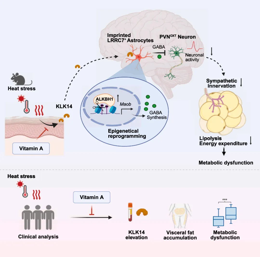
Study overview: heat stress activates a skin-derived KLK14 signal that imprints a persistent hypothalamic astrocyte state and alters downstream metabolic regulation.
The central biological observation was not that a single episode of heat directly caused obesity. Rather, prior heat stress altered the response to a later obesogenic challenge. Mice exposed to 37 °C for 6 hours per day over 7 days showed accelerated weight gain, impaired glucose tolerance, insulin resistance, and greater visceral adiposity after subsequent high-fat feeding.
The phenotype persisted for several weeks after heat exposure, supporting the authors' concept of a durable metabolic memory rather than a transient heat-shock response.
In parallel, analysis of 342 food-delivery workers in Changsha found that occupational exposure to temperatures of at least 37 °C was associated with higher BMI, waist circumference, visceral fat area, and HOMA-IR. Because this component was observational, it supports an association but does not by itself establish causality.
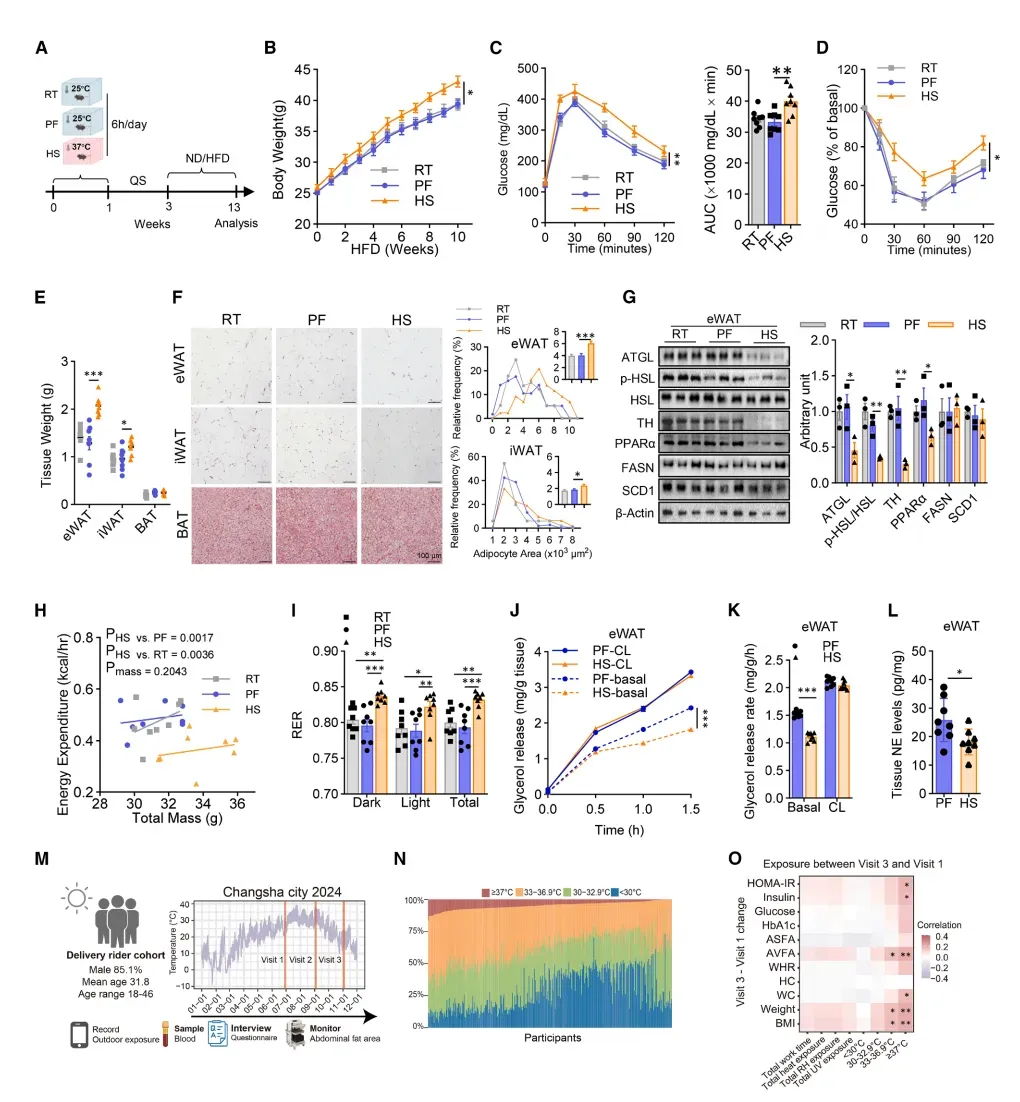
Figure 1. Prior heat stress increased susceptibility to diet-induced metabolic dysfunction in mice and was associated with adverse metabolic measures in a human occupational cohort.
Single-cell RNA sequencing resolved the response at cell-type level and identified a distinct LRRC7+ astrocyte population enriched in the paraventricular nucleus (PVN) after heat stress. This cell-state signal would have been difficult to distinguish in a bulk tissue average.
ATAC-seq revealed 3,338 regions with increased chromatin accessibility in these astrocytes. The accessibility program remained detectable for approximately 4 weeks and diminished by 8 weeks, closely matching the time course of the metabolic phenotype.
A second heat challenge rapidly reactivated the molecular program and produced a more pronounced metabolic response, providing functional support for a recallable, cell-specific heat memory.
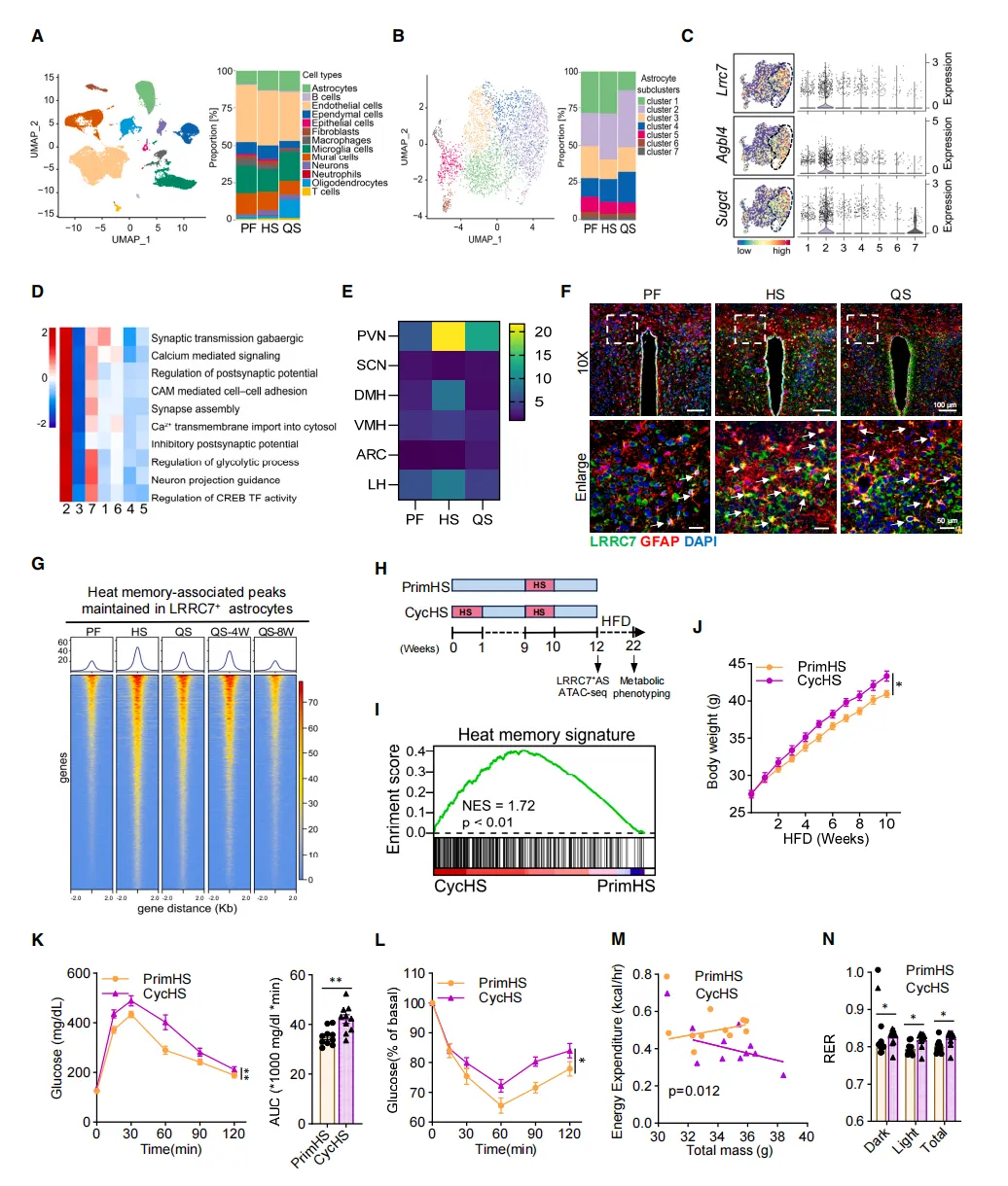
Figure 2. Single-cell and chromatin-accessibility analyses identify a persistent heat-associated program in LRRC7+ hypothalamic astrocytes.
The authors next traced the initiating signal to the skin. Heat stress increased KLK14 production in epidermal cells, raising circulating KLK14 and enabling signaling to the hypothalamus.
Genetic experiments showed that skin-specific loss of KLK14 prevented the persistent LRRC7+ astrocyte program and protected mice from the downstream metabolic phenotype. The study further identified CHL1 on astrocytes as the functional receptor linking circulating KLK14 to the hypothalamic response.
Together, these experiments move the proposed pathway beyond correlation: they connect a peripheral heat-sensing tissue to a defined central nervous system cell population through a specific ligand-receptor interaction.
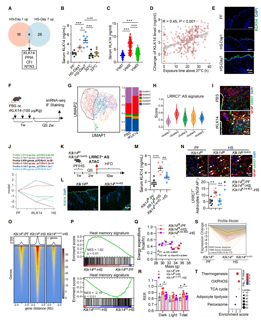
Figure 3. Skin-derived KLK14 mediates heat-induced imprinting of LRRC7+ hypothalamic astrocytes.
Mechanistically, KLK14-CHL1 signaling induced ALKBH1 in LRRC7+ astrocytes and reduced DNA N6-mA methylation. This change was accompanied by increased chromatin accessibility at loci associated with GABAergic synapses, calcium signaling, and related neural functions.
Perturbation of ALKBH1 disrupted the heat-memory signature and attenuated the downstream metabolic effects, positioning ALKBH1 as a key molecular bridge between extracellular KLK14 signaling and a durable astrocyte transcriptional state.
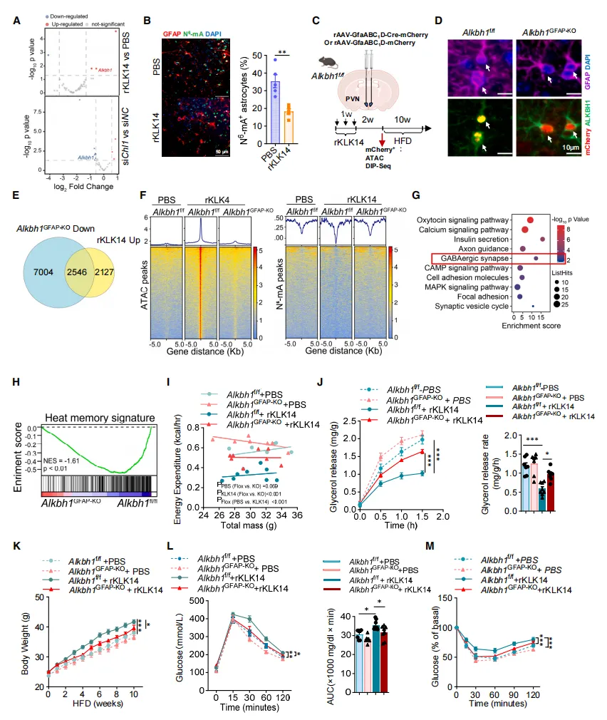
Figure 4. ALKBH1 regulates KLK14-induced epigenetic memory in LRRC7+ astrocytes.
The heat-imprinted LRRC7+ astrocytes increased GABA synthesis and inhibited oxytocin-expressing neurons in the PVN. This reduced sympathetic nervous system output to adipose tissue.
Lower sympathetic activity was accompanied by reduced norepinephrine signaling and decreased abundance or activation of key lipolytic proteins, including ATGL and phosphorylated HSL. The resulting reduction in fat mobilization favored visceral lipid accumulation during subsequent high-fat feeding.
This circuit-level evidence links a cell-specific epigenetic state in the hypothalamus to a measurable peripheral metabolic outcome.
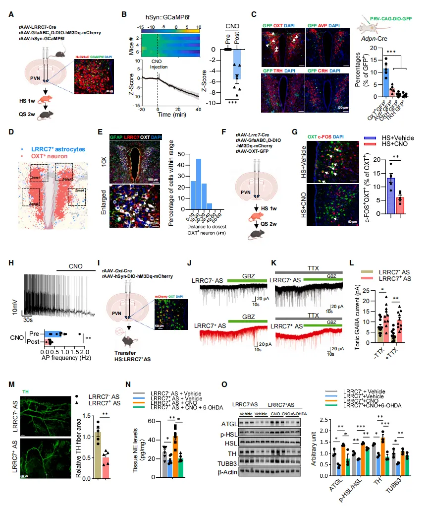
Figure 5. LRRC7+ astrocytes promote visceral fat deposition by suppressing PVN oxytocin neurons and sympathetic output to adipose tissue. Source: Zhou et al., Cell (2026).
The study linked heat exposure to reduced retinoid signaling in the skin and tested whether vitamin A could modify the KLK14 pathway. In mice, supplementation during heat exposure lowered KLK14 in skin and serum, reduced expansion of the LRRC7+ astrocyte population, and improved energy expenditure and lipolytic activity.
In the human intervention, participants received 5,000 IU of vitamin A per day or placebo for 8 weeks during heat exposure. Relative to placebo, vitamin A attenuated the rise in KLK14 and was associated with smaller increases in BMI, visceral fat area, and HOMA-IR at the dose and duration tested.
BMI increase: 5.04% with placebo versus 1.56% with vitamin A.
Increase in visceral fat area: 15.36 cm² lower in the vitamin A group.
Increase in HOMA-IR: 0.78 lower in the vitamin A group.
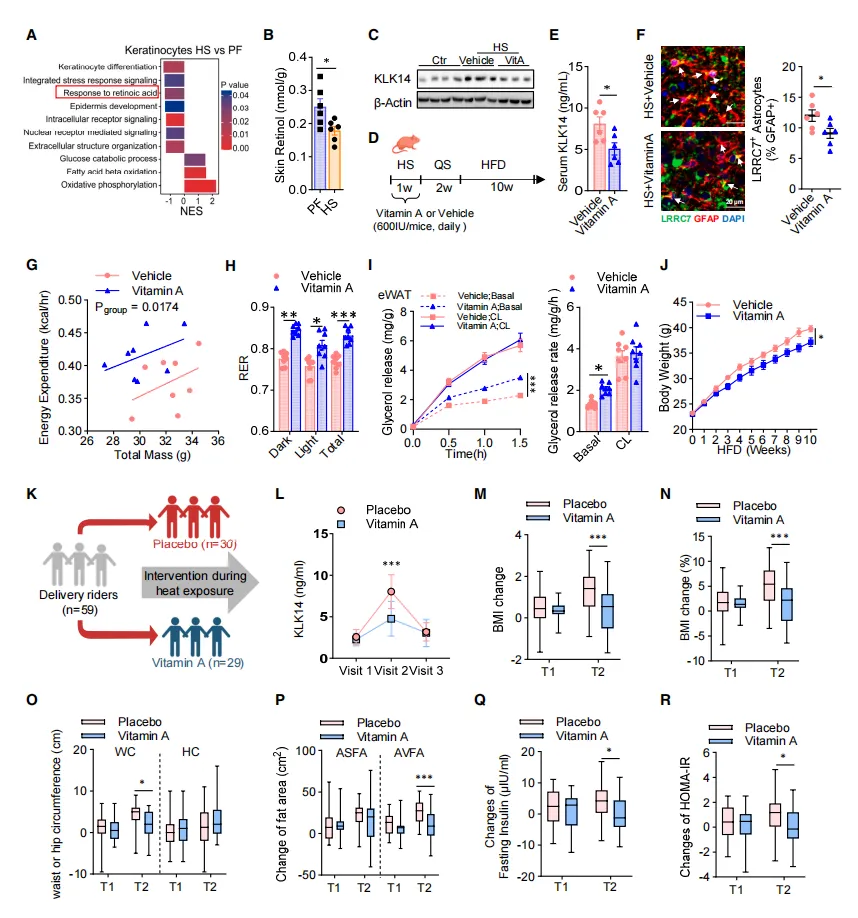
Figure 6. Vitamin A attenuated heat-associated KLK14 elevation and metabolic changes in mouse models and a short-term human intervention.
Resolved a heat-responsive LRRC7+ astrocyte population within a heterogeneous hypothalamic tissue.
Distinguished persistent cell-state changes from transient whole-tissue responses.
Provided a cellular anchor for ATAC-seq, ligand-receptor analysis, anatomical validation, and functional perturbation.
Supported a mechanistic model that could be tested across skin, brain, neural circuitry, and adipose tissue.
Omics Empower supports researchers with single-cell and single-nucleus RNA sequencing workflows, from sample feasibility assessment and experimental design to library preparation, sequencing, bioinformatics, and publication-ready data visualization.

Our team has supported more than 500 peer-reviewed publications across single-cell and spatial transcriptomics research, including studies published in Nature, Science, and Cell。
Whether you are working with fresh cells, fresh tissue, frozen samples, or clinically collected material, we can help assess the most suitable workflow for your project.
Zhou H-Y, Feng X, Wen J, et al. A skin-hypothalamus axis couples heat stress and metabolic dysfunction. Cell. 2026;189(12):3571-3588.e27. doi:10.1016/j.cell.2026.03.045.
scRNA-seq vs. snRNA-seq: How to Choose the Right Workflow for Your Stud
Comparing Single-Cell Sequencing Platforms: How to Choose the Right Fit for Your Study
A Complete Guide to Single-Nucleus RNA Sequencing (snRNA-seq)
Single-Cell Sequencing in Oncology Drug Development: Key Applications and Research Examples
Germany: Arnold-Graffi-Haus / D85 Robert-Rössle-Straße 10 13125 Berlin
United States: (CA) 2 Goddard, Irvine, CA 92618
United States: (IL) 8255 Lemont Rd, #1, Darien, IL 60561
Hong Kong: Unit 615, Building 11W, Hong Kong Science Park, Pak Shek Kok, Hong Kong
Germany: Arnold-Graffi-Haus / D85 Robert-Rössle-Straße 10 13125 Berlin
United States: (CA) 2 Goddard, Irvine, CA 92618
United States: (IL) 8255 Lemont Rd, #1, Darien, IL 60561
Hong Kong: Unit 615, Building 11W, Hong Kong Science Park, Pak Shek Kok, Hong Kong
Germany: Arnold-Graffi-Haus / D85 Robert-Rössle-Straße 10 13125 Berlin
United States: (CA) 2 Goddard, Irvine, CA 92618
United States: (IL) 8255 Lemont Rd, #1, Darien, IL 60561
Hong Kong: Unit 615, Building 11W, Hong Kong Science Park, Pak Shek Kok, Hong Kong

 EN
EN
 de
de